01. AUTOMATED ORGAN SEGMENTATION CORRECTION


Automated tools can segment organs and muscles quickly, and the result can look impressively clean in 3D. Smooth surfaces, plausible shapes, nothing obviously wrong to the untrained eye. But errors often hide at boundaries with neighbouring structures: over- or under-segmentation that looks minor but skews volumes, lesion quantification or radiomics.
The example below shows a nice-looking 3D segmentation, but looking at the 2D native images, I quickly see multiple errors: undersegmentation of the left iliopsoas, the gluteus maximus, the deep back muscles, the bladder and the small bowel. These are easy to miss on a quick visual check, but significant once the data is used downstream.
I review and correct automated output from your propriety tool, or from a range of open-source tools like the one in the example, bringing it to a clinically and scientifically reliable standard. Corrected segmentations support quantitative research (volumes, imaging biomarkers, radiomics) and clinical use: i.e., accurate liver and segmental volumes matter directly for surgical planning. Depending on the project, I can also build a segmentation from scratch, with a treating clinician's review as a final check where appropriate.
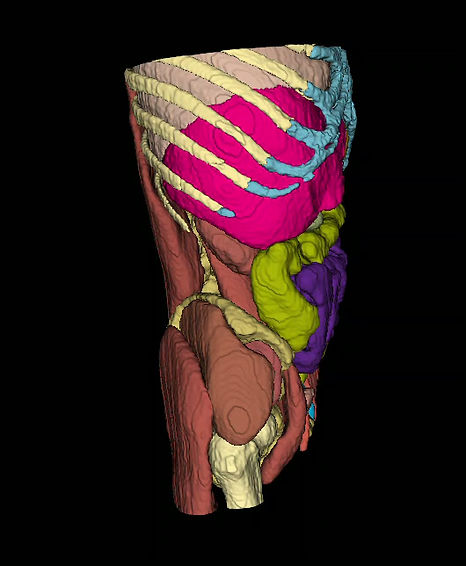

3D visualisation vs. native images: undersegmentation of the left iliopsoas, gluteus maximus, deep back muscles, bladder and small bowel by automated algorithm.
02. BRAIN SEGMENTATION


QC reports from modern neuroimaging pipelines can look reassuring while missing real anatomical errors. Tissue segmentation can wrongly include structures like the dura, falx or tentorium, or oversegment grey matter with poorly finished boundaries, particularly in the frontal and temporal lobes (see example above) and around the skull base. These errors rarely show up in standard metrics but can skew downstream measurements.
I review and correct output from open-source or proprietary brain segmentation tools, refining boundaries and removing misclassified structures against the native scan. Often the pipeline is already largely correct. The work is fixing the last few percent that matters. How much correction is needed depends on the application: large-cohort studies can tolerate errors that individual-level or clinical analysis can't.