01. INTRACRANIAL PATHOLOGY SEGMENTATION


Traumatic brain injury is one of the hardest segmentation problems in imaging: injuries are often multifocal, heterogeneous and poorly defined, with more than 20 lesion types recognised by the National Institute of Neurological Disorders and Stroke (NINDS). Automated tools have improved, but progress is limited by a shortage of high-quality, voxel-level ground-truth data. Even experienced specialists disagree on where boundaries lie, especially for subarachnoid haemorrhage, early oedema and subtler injuries.
I've manually segmented hundreds of CT and MRI studies over 10+ years, including work on an AI system that went on to receive CE marking and FDA clearance. In my current academic research I use multi-expert annotation and consensus methods (e.g. STAPLE) to build more reliable ground truth, with SOPs developed for consistent segmentation of complex pathology in 3D Slicer.
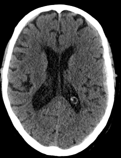


Because TBI has forced me to confront some of the hardest segmentation problems in medical imaging, I've been able to carry that same rigor into other anatomical areas.
02. VISCERAL ANATOMY SEGMENTATION

Thoracic, abdominal and pelvic organs are usually easier to segment than TBI, but not automatic: anatomy varies between individuals, and boundary visibility depends on modality, contrast and surrounding structures. Pathology adds further complexity.
I manually segment organs, vessels, musculature and other structures, adapting the approach to the imaging and the question at hand, whether that's simply organ volume, or a lesion mapped alongside nearby vessels to show their 3D relationship. Segmentations can also be reconstructed in 3D for surgical planning, research or printing. Different projects need different levels of detail, and I tailor the work accordingly.